
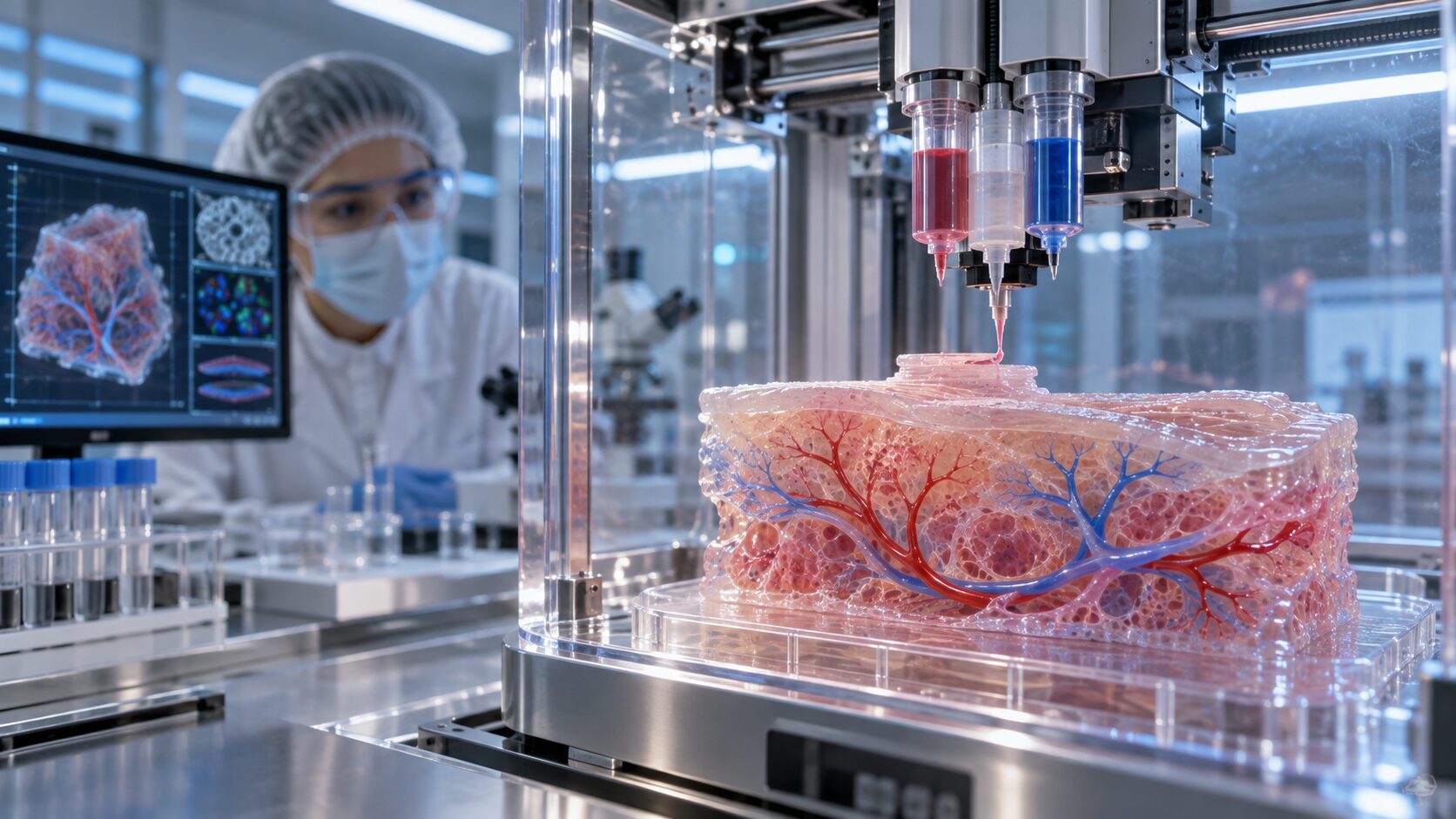
Printing Human Tissue Is No Longer Science Fiction—So What Is Bioprinting Still Waiting For?
The idea sounds almost impossible: load living cells into a machine, arrange them layer by layer and print replacement tissue for the human body.
Yet researchers are already bioprinting skin-like structures, cartilage, bone scaffolds, blood vessels and miniature models of organs. These constructs are helping scientists study disease, test drugs and explore personalized treatments. NASA has even investigated bioprinting in microgravity, where fragile biological structures can potentially be created without fighting Earth’s gravity. NASA
So why can’t a hospital simply print a new kidney, heart or liver for a patient?
The short answer is that printing the shape of an organ is not the same as reproducing its biology.
The Blood-Vessel Problem
The biggest obstacle is vascularization—the creation of a working network of blood vessels.
Cells need a continuous supply of oxygen and nutrients. In thin tissues, those materials can diffuse across short distances. In a thick organ, cells buried deep inside the structure will quickly become starved unless blood can reach them.
A printed kidney or liver would therefore need an extraordinarily detailed network of large vessels, tiny capillaries and microscopic connections. Those vessels must remain open, connect with the patient’s circulation and respond naturally to blood pressure.
Until that vascular network can be printed reliably, keeping a full-sized engineered organ alive remains extremely difficult.
Keeping Cells Alive During Printing
Living cells are far more delicate than plastic or metal.
During bioprinting, they may be exposed to pressure, shear forces, temperature changes, ultraviolet light or chemical crosslinking. Researchers must deposit cells precisely without damaging them—and then provide an environment in which they can survive, mature and begin working together.
This creates a difficult balancing act. A bioink must be soft enough to protect cells but strong enough to retain its printed shape. It must also allow nutrients to circulate and cells to communicate.
A structure that looks like tissue under a microscope is not necessarily functional tissue. Heart cells must contract together. Nerve cells must transmit signals. Liver cells must process chemicals. Kidney cells must filter waste. Recreating those coordinated behaviors is much harder than printing the correct outline.
Biology Does Not Behave Like a Factory
Even if researchers produce a successful tissue once, medicine requires them to produce it repeatedly.
Hospitals and regulators need assurance that every printed product contains the correct cells, maintains its strength, remains sterile and performs predictably. Small differences in a patient’s cells, the bioink, printing pressure or maturation process can change the final result.
Scaling production will require standardized materials, automated quality control and systems capable of monitoring tissue as it develops. The industry must move from impressive laboratory demonstrations to repeatable medical manufacturing.
That is a much higher standard.
Regulation Must Evaluate the Entire Process
A bioprinted implant may combine living cells, biomaterials, biological signals and a digitally manufactured structure. That makes it more complicated to evaluate than a conventional drug or medical device.
Regulators must consider where the cells came from, how they were modified, whether the product could cause tumors or immune reactions, how it was manufactured and whether it remains safe after implantation. In the United States, some tissue-engineered products may fall within the FDA’s regenerative-medicine framework, including pathways intended to accelerate qualifying therapies for serious conditions. FDA
The challenge is not that regulation is preventing innovation. It is that living, patient-specific products create questions traditional approval systems were not originally designed to answer.
What Will Reach Patients First?
The first widely used bioprinted products probably will not be complete hearts or kidneys.
Simpler, thinner and less vascular tissues have a more realistic path forward. These may include:
- Skin for burns and wound repair
- Cartilage for joint or facial reconstruction
- Bone-repair scaffolds
- Corneal and other thin tissue structures
- Small vascular grafts
- Tissue patches that support a damaged heart or liver
Bioprinted tissue models used outside the body could advance even faster. Pharmaceutical companies can use miniature liver, tumor, intestinal and cardiac tissues to test drug toxicity and effectiveness. Patient-derived cells could eventually help doctors identify which treatment is most likely to work before exposing the patient to it.
These research applications do not require a complete transplantable organ to deliver enormous value.
The Real Bioprinting Revolution
The future of bioprinting is often illustrated by a machine producing a perfect human heart. That day may come, but it will not arrive through one spectacular breakthrough.
It will emerge through steady progress in vascularization, stem-cell biology, bioinks, automation, imaging, artificial intelligence and manufacturing standards.
Bioprinting is no longer waiting for proof that human tissue can be printed. That question has largely been answered.
It is waiting for printed tissue to stay alive, mature correctly, perform reliably and be manufactured safely at medical scale.
The revolution has already started—it is simply beginning with tissue models, repair patches and smaller structures before progressing toward complete replacement organs.
Leave a Reply